Ed Friedlander, M.D., Pathologist
scalpel_blade@yahoo.com
No texting or chat messages, please. Ordinary e-mails are welcome.
|

|
 |
 |
 |
 |
|
verify here. |
Cyberfriends: The help you're looking for is probably here.
This website collects no information. If you e-mail me, neither your e-mail address nor any other information will ever be passed on to any third party, unless required by law.
This page was last modified January 1, 2016.
I have no sponsors and do not host paid advertisements. All external links are provided freely to sites that I believe my visitors will find helpful.
Welcome to Ed's Pathology Notes, placed here originally for the convenience of medical students at my school. You need to check the accuracy of any information, from any source, against other credible sources. I cannot diagnose or treat over the web, I cannot comment on the health care you have already received, and these notes cannot substitute for your own doctor's care. I am good at helping people find resources and answers. If you need me, send me an E-mail at scalpel_blade@yahoo.com Your confidentiality is completely respected. No texting or chat messages, please. Ordinary e-mails are welcome.
 I am active in HealthTap,
which provides free medical guidance from your cell phone.
There is also a fee site at
www.afraidtoask.com.
I am active in HealthTap,
which provides free medical guidance from your cell phone.
There is also a fee site at
www.afraidtoask.com.
 If you have a Second Life account, please visit my teammates and me at the Medical Examiner's office. |
|
 |
With one of four large boxes of "Pathguy" replies. |
 I'm still doing my best to answer
everybody.
Sometimes I get backlogged,
sometimes my E-mail crashes, and sometimes my
literature search software crashes. If you've not heard
from me in a week, post me again. I send my most
challenging questions to the medical student pathology
interest group, minus the name, but with your E-mail
where you can receive a reply.
I'm still doing my best to answer
everybody.
Sometimes I get backlogged,
sometimes my E-mail crashes, and sometimes my
literature search software crashes. If you've not heard
from me in a week, post me again. I send my most
challenging questions to the medical student pathology
interest group, minus the name, but with your E-mail
where you can receive a reply.
Numbers in {curly braces} are from the magnificent Slice of Life videodisk. No medical student should be without access to this wonderful resource.
 I am presently adding clickable links to
images in these notes. Let me know about good online
sources in addition to these:
I am presently adding clickable links to
images in these notes. Let me know about good online
sources in addition to these:
 My team:
pathology.org -- my cyberfriends, great for current news and browsing for the general public
EnjoyPath -- a great resource for everyone, from beginning medical students to pathologists with years of experience
Medmark Pathology -- massive listing of pathology sites
Estimating the Time of Death -- computer program right on a webpage
Pathology Field Guide -- recognizing anatomic lesions, no pictures
Freely have you received, freely give. -- Matthew 10:8. My site receives an enormous amount of traffic, and I'm still handling dozens of requests for information weekly, all as a public service.
Pathology's modern founder, Rudolf Virchow M.D., left a legacy of realism and social conscience for the discipline. I am a mainstream Christian, a man of science, and a proponent of common sense and common kindness. I am an outspoken enemy of all the make-believe and bunk that interfere with peoples' health, reasonable freedom, and happiness. I talk and write straight, and without apology.
Throughout these notes, I am speaking only for myself, and not for any employer, organization, or associate.
Special thanks to my friend and colleague, Charles Wheeler M.D., pathologist and former Kansas City mayor. Thanks also to the real Patch Adams M.D., who wrote me encouragement when we were both beginning our unusual medical careers.
If you're a private individual who's enjoyed this site, and want to say, "Thank you, Ed!", then what I'd like best is a contribution to the Episcopalian home for abandoned, neglected, and abused kids in Nevada:

My home page
More of my notes
My medical students
Especially if you're looking for information on a disease with a name that you know, here are a couple of great places for you to go right now and use Medline, which will allow you to find every relevant current scientific publication. You owe it to yourself to learn to use this invaluable internet resource. Not only will you find some information immediately, but you'll have references to journal articles that you can obtain by interlibrary loan, plus the names of the world's foremost experts and their institutions.
Alternative (complementary) medicine has made real progress since my generally-unfavorable 1983 review. If you are interested in complementary medicine, then I would urge you to visit my new Alternative Medicine page. If you are looking for something on complementary medicine, please go first to the American Association of Naturopathic Physicians. And for your enjoyment... here are some of my old pathology exams for medical school undergraduates.
I cannot examine every claim that my correspondents
share with me. Sometimes the independent thinkers
prove to be correct, and paradigms shift as a result.
You also know that extraordinary claims require
extraordinary evidence. When a discovery proves to
square with the observable world, scientists make
reputations by confirming it, and corporations
are soon making profits from it. When a
decades-old claim by a "persecuted genius"
finds no acceptance from mainstream science,
it probably failed some basic experimental tests designed
to eliminate self-deception. If you ask me about
something like this, I will simply invite you to
do some tests yourself, perhaps as a high-school
science project. Who knows? Perhaps
it'll be you who makes the next great discovery!
Our world is full of people who have found peace, fulfillment, and friendship
by suspending their own reasoning and
simply accepting a single authority that seems wise and good.
I've learned that they leave the movements when, and only when, they
discover they have been maliciously deceived.
In the meantime, nothing that I can say or do will
convince such people that I am a decent human being. I no longer
answer my crank mail.
This site is my hobby, and I do not accept donations, though I appreciate those who have offered to help.
During the eighteen years my site has been online, it's proved to be one of the most popular of all internet sites for undergraduate physician and allied-health education. It is so well-known that I'm not worried about borrowers. I never refuse requests from colleagues for permission to adapt or duplicate it for their own courses... and many do. So, fellow-teachers, help yourselves. Don't sell it for a profit, don't use it for a bad purpose, and at some time in your course, mention me as author and William Carey as my institution. Drop me a note about your successes. And special thanks to everyone who's helped and encouraged me, and especially the people at William Carey for making it still possible, and my teaching assistants over the years.
Whatever you're looking for on the web, I hope you find it, here or elsewhere. Health and friendship!

![]()
![]()
Review the major players in the immune system, both cellular and humoral. Be sure you can explain what a hapten is.
For each of the following, describe the essential mechanism and mention some real-life illustrations: Type I, Type II, Type III, and Type IV hypersensitivity injury.
Briefly describe each of the following:
common hay fever / asthma
urticaria
systemic anaphylaxis
food allergy
Type II (fixed antigens)
Complement-mediated
ABO-mismatched transfusion reaction
Rh-mismatched delayed transfusion reaction
Autoimmune hemolytic anemia
Autoimmune thrombocytopenia
Goodpasture's disease of the lung and kidney
Pemphigus family of skin diseases
Hyperacute and acute transplant rejection (also some type III)
Cell-mediated ("antibody-dependent cell-mediated cytotoxicity")
Antibody-mediated dysfunction
circulating anticoagulants
excitatory and inhibitory antibodies (Graves', many others)
Type III (immune complex disease; soluble antigens)
Arthus reaction -- serum sickness
aches and pains of the "'flu"
Immune-complex induced thrombocytopenia (AIDS, idiopathic)
vasculitis (hepatitis B, rheumatoid arthritis)
Type IV (delayed hypersensitivity, generally without antibody involvement)
tuberculin skin test
nickel skin allergy
acute cellular transplant rejection
graft-versus-host diseases
"anergy" (explain the idea)
Describe and recognize the various anatomic signs of transplant rejection and graft versus host disease.
Critique the following statement, overheard at a party: "By strengthening our immune system, we prevent or cure most diseases. We strengthen our immune system by nutritional supplements and mental imagery to reduce stress."
|
|
|
![]() KCUMB Students
KCUMB Students
"Big Robbins" -- Immuno
Lectures follow Textbook
QUIZBANK
Immuno (all; this covers these 4 lectures)
Metabolic (amyloid) #'s 93-111
Clinical immunology lab (all)
LEARN FIRST: TYPES OF IMMUNE INJURY ("HYPERSENSITIVITY")
Okay. These divisions are artificial.
Type I... Havoc resulting from IgE and mast-cell degranulation
Type II... Antibodies bind to cells. This can cause
Type III... Antigen-antibody complexes ("immune complexes") precipitate and cause the damage
Type IV... Havoc wrought by specifically-sensitized T-cells without involvement of antibodies. This includes...
CLASSIC DELAYED HYPERSENSITIVITY, in which the harm is done primarily by recruited macrophages and innocent bystander cells are also massacred. No antibody is involved.
ANTIBODY-INDEPENDENT CELL-MEDIATED CYTOTOXICITY, in which individual cells are assassinated ("apoptosis") by T-CTL lymphocytes, without harm to innocent bystanders. This term is still in some use.
Exactly which illnesses generally believed to be T-cell initiated will end up in this category remains speculative.
No one uses the term for such illnesses as most viral hepatitis, in which most of the damage is done by T-cells removing infected cells.
HAPTENS are atoms or small molecules that elicit an immune response when, and only when, they are bound to a larger carrier molecule, generally a protein. The body's rejection of the hapten-protein adduct does the damage. Some famous examples are:
Many (maybe most) really surprising, rare idiosyncratic drug reactions involve the drug acting as a hapten.
CELLS OF THE IMMUNE SYSTEM
|
{14252} lymphocytes
|  Osmosis Jones |
* Click here for a video-game in which you are the immune system fighting pseudomonas bacteria.
T-LYMPHOCYTES (T-cells):
A versatile family of cells that have passed through the thymus (and been acted on by the hormone thymosin) during early biological life.
T-cells are the center-piece of the immune system. T-cells of various sorts are predominant throughout the lymph nodes except in and around germinal centers. They also make up most of the white pulp of the spleen, and occupy the thymus gland. Around 80% of circulating lymphocytes are T-cells.
Young T-cells differentiate into many subtypes, which act as regulators and effectors.
Each T-cell acquires its specificity, which it transmits to its descendants, by recombination of its TCR gene before birth.
CD3 -- joins with the TCR surface molecule, but is not rearranged. General T-cell marker.
CD4 -- helper cell marker
CD8 -- killer cell marker (cytolytic T's, natural killers)
CD28 -- TCR co-receptor, marker for T-cells
T-helper cells (TH-cells) activate B- and other T-cells
Many subclasses exist to do different things. You'll learn about them in "Immunology".
Worth remembering: TH1's produce IL-2 and INF-gamma and act on macrophages; TH2's produce IL-4 and IL-5 and work on the eosinophils.
T-cytolytic (T-CTL-cells, T-killer cells) cells attach to and kill (apoptosis) cells bearing whatever antigen against which they are specifically programmed and sensitized. These cells may be T4+ or T8+.
After a T-cell has responded to a particular antigen and proliferated, some members of the clone survive and thus the response to the antigen is stronger next time it is encountered ("clonal expansion").
B-LYMPHOCYTES (B-cells):
Another class of lymphocytes, which when activated produce freely circulating immunoglobulins ("antibodies", "gamma globulins", "humoral immunity").
Antibodies coat harmful foreign particles, and bind to foreign molecules.
Sometimes this helps (pneumococcal pneumonia, measles![]() , hepatitis B).
, hepatitis B).
Sometimes it doesn't seem to help (AIDS, many other infections, cancer).
Sometimes it hurts (ragweed allergy, autoimmune hemolytic anemia, systemic lupus, pernicious anemia, and every other type I, type II, and type III hypersensitivity disease).
B-cells predominate in the follicles (germinal centers) of the lymph nodes and are more common than T-cells in the red pulp of the spleen and in the bone marrow.
In the germinal centers, antigen-antibody complexes are presented to baby B-cells by the dendritic follicular cells (seen 'em?); the ones that match the antigen best are caused to divide and mature (Nature 367: 425, 1994). These will be the ones that make IgG.
Around 15% of circulating lymphocytes in the peripheral blood are B-cells.
B-cells have surface immunoglobulin, which is how we usually spot them.
There are a host of other markers that recoginze B-cells in various stages of differentiation. Don't worry about these.
B-cell activation is partially understood.
Before birth, each B-cell gets its immunoglobulin gene rearranged to recognize a particular antigen. The antibody that the B-cell will make is displayed on its surface, with its business end pointing straight outward.
If and when this antigen comes along, it triggers the B-cell, which starts dividing (under the modulation of T-cells, of course).
After assuming a variety of curious shapes (look at the cells within a germinal center), most of the original B-cell's progeny become a "selected clone" of plasma cells pumping out specific antibody.
{46429} plasma cell, electron micrograph
When the immune system wins, the plasma cells mostly die, but plenty of the original B-cell's clones survive and "remember". Next time the antigen comes along, the antibody response will be much greater ("anamnestic response", i.e., "we haven't forgotten").
Remember: you can't tell resting B-cells from resting T-cells by their morphology.
NATURAL KILLER LYMPHOCYTES:
"Natural killer lymphocytes" (NK-cells) are bigger than other lymphocytes, and have cytoplasmic granules (i.e., you can pick them out on a smear; they make up around 10% of the circulating cells).
They attack and cause apoptosis in cells with altered class I MCH antigens, without being "specially sensitized".
The granules, which are injected into the target cell, contain perforin and granzyme, along with other stuff (* TIA-1 etc.).
CD57 -- cytotoxic natural killers
MONONUCLEAR PHAGOCYTES
This is all of the "reticuloendothelial system", found throughout the body. Probably this is the oldest component of the immune system.
The familiar wandering macrophages and microglia are monocytes from the peripheral blood that have entered the tissues. (Same cell, different names.)
Fixed phagocytes (the rest of the "mononuclear phagocytic system") line the vascular channels of the body. They, too, were once circulating monocytes.
A special subclass of mononuclear phagocytes, the "dendritic" and "Langerhans'" cells (together, "the accessory cells", etc.), seem to be the best antigen processors and the poorest effector phagocytes. They have special surface antigens (Ia -- Immune activation antigens coded by HLA genes) and have other special features.
Mononuclear phagocytes are essential to immune responses.
The "accessory cells" ("dendritic" and "Langerhans'") apparently phagocytize antigens, process them (into "epitopes"), attach them to Ia antigens, and present them to lymphocytes to begin the immune response.
Busy macrophages modulate the activities of other cells by secreting soluble factors such as interleukin 1 and TNF-alpha (see above), as well as some complement components (supplementing the bulk, which come from the liver).
In addition, macrophages are effector cells when summoned by T-cells or activated complement. They devour and sometimes digest harmful things, and can become epithelioid cells of granulomas to wall off other bad things.
They have receptors for the Fc portion of IgG and for various complement components.
The milieu of sex hormones has a host of effects on immune function, and you will be completely mystified if you try to sort through even a few papers. If you are going to approach this subject at all, you may enjoy J. Leuk. Bio. 75: 1166, 2004 and J. Imm. 175: 5716, 2005, both first-authored by our own Chad Lambert '09.
We stain for common macrophages using CD68. We stain for antigen-presenters with CD1a.
{08230} macrophage containing
leishmania![]() (Baghdad boil)
(Baghdad boil)
MAST CELLS AND BASOPHILS
These cells are rich in vasoactive (and other) substances. In addition to the familiar histamine (right away) and leukotrienes (after a while), remember endothelium relaxing factor and neutrophil chemotactic factor.
When mast cells are activated (usually by something reacting with the IgE that covers their surfaces), the substances are released (histamine right away, leukotrienes over a few hours). These have a variety of effects on blood vessels, smooth muscle, gastric parietal cells, etc.
* In 1989, at a scientific meeting, the author got into it with French biochemist Jacques Benveniste, who claimed to have demonstrated an effect of homeopathic-type dilutions on basophil degranulation. Dr. Benveniste showed a phase contrast photo that the author believed was a neutrophil, and called it a degranulated basophil. Dr. Benveniste graciously acknowledged that making the distinction was difficult. I believe he is sincere, if perhaps wrong. For more on Dr. Benveniste, see Skeptical Inquirer 22(1): 19, Jan-Feb 1998.
{09205} mast cell granules (electron micrograph)
{13733} mast cells
{14539} mast cells (purple ones) and fibroblasts
{14542} mast cell degranulating
{15117} mast cell city!
{46472} mast cell, electron micrograph, intact
{46473} mast cell, electron micrograph, degranulating
NEUTROPHILS: You are already familiar with these important commando cells. They continue to surprise -- one recent review is Chest 134: 606, 2008.
Polys elaborate at least 50 distinct destructive substances. Remember that, while they fight infection, neutrophils also do tremendous damage to the body itself.
In the common (1/2000 people) "chronic granulomatous disease", the neutrophils cannot produce
peroxide to kill staphylococci![]() ,
so the epithelioid macrophages must take over (hence the name of the
disease).
,
so the epithelioid macrophages must take over (hence the name of the
disease).
EOSINOPHILS: Review in J. Allerg. Clin. Immuno. 105: 651, 2000.
{09207} eosinophil granules
{14099} eosinophils
{14708} eosinophil
IMMUNOGLOBULINS and COMPLEMENT should be familiar to you. For a pathologist's perspective on complement today, see Am. J. Path. 171: 715, 2008.
IgM (our most important macroglobulin) molecules are large pentamers, tend to be cold-reactive, appear early during the humoral immune response, and can fix complement when just one molecule binds to something. IgG molecules are usually monomers, tend to be warm-reactive, appear later during humoral response, include some classes that can fix complement when two or more molecules find to something, and comes from B-cells clones with good memories. IgA molecules tend to be found on mucosal surfaces and in mothers' milk. IgD is a receptor on a B-cell, and its significance in the circulating blood remains obscure. IgE sticks by its Fc portion to basophils and mast cells, ready to tell them to degranulate.
Early in the 21st century, the "IgG4 related disease" group was identified. Cartwheel ("storiform") fibrosis (except in lymph nodes)and fibrotic, inflamed medium-sized veins are the hallmarks regardless of location, along with elevated blood IgG4 levels and lots of IgG4 plasma cells (non-clonal, NEJM 366: 539, 2012). Facial lesions (Am. J. Surg. Path. 37: 66, 2013) and sclerosing pancreatitis are only the best-known features; probably every organ can be affected. Not well understood, it responds to rituximab, and perhaps thalidomide (Jama Derm 149: 742, 2013).
Idiotypes are the specific recognition sequences that make a particular antibody unique. Watch for "anti-idiotype" molecules as an up-and-coming way of modulating the immune system (and evidence that it's one way in which we modulate our own system. Note that an anti-idiotype antibody is likely to resemble the antigen, since both bind to the same location.)
The complement cascade, when activated, increases vascular permeability, summons polys and monos, and punches holes in membranes. All chordates have complement.
CYTOKINES
Immune cells send messages by cytokines (formerly called "lymphokines"), and receive similar messages from other cells. Over a dozen of these "biologic response modifiers" are well-characterized, and several are now undergoing clinical trials.
INTERLEUKIN 1: two "monokines" produced in bulk by macrophages that are phagocytizing or hurting, this substance causes T-cells to multiply. It also causes the acute-phase reaction (* with interleukin 6 and TNF; you will learn about the acute phase reaction later), acts on the brain to cause fever and fatigue (etc., key paper Science 240: 321, 1988), causes catabolism of muscle in acute illness, recruits macrophages for granuloma formation, and is largely responsible for the increase in circulating neutrophils.
* Interleukin-1 receptor antagonist (IL-1RA; "anakinra" etc.) is yet another molecule in your internal milieu. For years these notes mentioned that this is a molecule in search of a disease for which it's the magic bullet. Today, the rare "neonatal-onset multisystem inflammatory disease", with a lack of cryopyrin (CIAS1 locus), is more mangeable with anakinra (NEJM 355: 581, 2006); it's also proved useful in refractory rheumatoid arthritis.
INTERLEUKIN 2 (IL-2, "T-cell growth factor"): a lymphokine from activated T4+ cells that promotes the division of other T-cells.
This -- with or without killer cells -- is a perennial experimental cancer therapy. It causes severe fatigue and insanity.... (horror stories Ann. Int. Med. 107: 293, 1987), and old reports that it was extremely elevated in the controversial "chronic fatigue syndrome" ("myalgic encephalomyelitis"; "Yuppie 'flu", Ann. Int. Med. 110: 321, 1989) have failed to find additional support; this claim (and others about cytokines, in chronic fatigue syndrome and in fibromyalgia) have failed by today's tests (J. Clin. Immuno. 19: 414, 1999; Clin. Exp. Imm. 132: 360, 2003). Whether there is some more subtle disturbance remains unknown -- I still suspect there is.
Its use in treating kidney cancer is one of the few immune-strengthing intervention that actually works, sort-of, sometimes: Science 255: 528, 1992. Possibly the archaic bacterially-derived "Coley's toxins" worked against cancer by making the body produce interleukins (Nature 357: 11, 1992).
IL-2 receptor levels, from breakdown of T cells, lets you know level of activity of immune disease.
The interleukin 2 receptor is now the target for a variety of therapeutic monoclonal antibodies, including daclizumab ("Zenapax", now in use for multiple sclerosis, psoriasis, ulcerative colitis, and other illnesses)
INTERLEUKIN 5 is the great mobilizer of eosinophils: NEJM 324: 1110, 1991 (still good). It is blocked by meprolizumab, which has shown great promise in managing eosinophil-related diseases such as severe asthma (NEJM 360: 973, 2009).
INTERLEUKIN 8 is chemotactic for neutrophils.
INTERFERONS: a class of substances, some produced by T-cells, which "interfere with viruses" and have a huge variety of other activities.
ALPHA (alfa) INTERFERON (* many subtypes exist) is a good therapy for hairy-cell leukemia, chronic hepatitis B and C, essential thrombocythemia, laryngeal warts, chronic granulocytic leukemia (nobody knows how this works), and "chronic granulomatous disease" (i.e., weak polys). Successes against cancer have been less impressive, though it's helped some melanoma patients. Administration causes a 'flu-like syndrome; it also acts on the brain, causing severe fatigue and/or even insanity.
BETA INTERFERON is the old name for B-cell stimulating factor, which turns B-cells into plasma cells. Two forms are in use (IFBeta-1a and IFBeta-1b), an important part of the treatment of multiple sclerosis.
GAMMA INTERFERON seems to be the principal substance that makes macrophages angry and form
granulomas, and the substance that causes macrophages to express Ia antigens.
Clinical uses now include leprosy![]() ,
leishmaniasis
,
leishmaniasis![]() ,
atypical mycobacteria,
and stubborn venereal warts.
It can be administered by vein (the 'flu-like syndrome it produces is mild), or inhaled for lung disease.
,
atypical mycobacteria,
and stubborn venereal warts.
It can be administered by vein (the 'flu-like syndrome it produces is mild), or inhaled for lung disease.
Someday we may inhibit interferon-gamma in order to prevent intestinal adhesions after surgery (perhaps using hepatocyte growth factor: Nat. Med. 14: 437, 2008).
TUMOR NECROSIS FACTOR: a "monokine" ("tumor necrosis factor alpha", "cachectin", now usually called simply "tumor necrosis factor" or "TNF") and a closely-related "lymphokine" (tumor necrosis factor beta", now usually called simply "lymphotoxin"), tandem homologous gene products that mediate a wide range of tissue injuries.
* This was a hot topic especially in the 1990's. Everything from endotoxic shock and "the Schwartzman reaction" (an old experimentalist's disease model with two doses of endotoxin, the second proving fatal -- historic interst) to the Herxheimer response (sickness after syphilis treatment due to the dead spirochetes -- NEJM 335: 311, 1996) to Waterhouse-Friderichsen syndrome to extreme over-activation of arachidonate metabolite systems to cachexia of cancer and AIDS is attributed to these substances. Key review NEJM 316: 379, 1987; still good. TNF causes emaciation by blocking adipocyte lipoprotein lipase -- does anyone want to try it for dieters?
* Early in the AIDS epidemic, we discovered that TNF also participates in granuloma formation, probably acting as an autocrine factor (Cell 56: 731, 1989, Clin. Imm. Imm. 51: 419, 1989). TNF is able to generate granulomas in the absence of any lymphocytes (Nature 356: 604, 1992), contradicting classic assumptions ("All granulomas are held together by gamma interferon"), and explaining why AIDS patients can still make reasonable granulomas.
Thalidomide selectively inhibits alpha-TNF production (Proc. Nat. Acad. Sci. 90: 5974, 1993 was key) and is finding uses in clinical medicine (sarcoidosis, inflammatory bowel disease, ankylosing spondylitis, Behcet's, recalcitrant aphthae in AIDS, even congestive heart failure).
* In the mid-1990's, a group of clinicians tried to help septic shock patients by removing tumor necrosis factor using antibodies. This didn't help, and seems to have killed a bunch of people, probably by a type III immune mechanism: NEJM 334: 1697, 1996.
TNF receptors are a story in themselves, and include the apoptosis equipment (no surprise): NEJM 334: 1717, 1996. One of the great success stories of today's medicine is administration of recombinant TNF receptors to soak up TNF itself in various diseases.
TRANSFORMING GROWTH FACTOR BETA exists as isoforms that apparently turn down most aspects of chronic inflammation. Mice born without the first isoform die young of widespread chronic inflammation (Proc. Nat. Acad. Sci. 90: 770, 1993).
The CHEMOKINES bring in and (sometimes) activate specific subsets of white cells, etc., etc. Lancet 349, 1997.
Simply injecting the DNA for a particular protein into muscle or dermis commonly results in B-cell and T-cell immunity to that protein (Proc. Nat. Acad. Sci. 91: 9519, 1994). Despite much excitement, the response isn't good enough to compare with real vaccines presently in use.
TYPE I IMMUNE INJURY IN HUMAN DISEASE
Type I hypersensitivity ("anaphylactic" or "immediate hypersensitivity") is the result of antigen binding to IgE on the surface of mast cells and basophils. These instantly degranulate and release active substances into the surrounding tissue.
The reaction begins within seconds to minutes following antigen exposure, and is over within a few hours.
IgE protects us against worms. (Almost everyone becomes allergic to ascaris worm once they meet it.) The idea that type I hypersensitivity was created to help protect us from worms has found good experimental support: Nature 349: 243, 1991.
* IgE production is of course orchestrated by T-helper and T-suppressor cells (T-suppressors are now "Treg cells") that specialize in just this.
* A few unusual types of IgG attached to mast cells can do exactly the same thing that IgE does.
Some people make IgE much more readily than others, and these are the people with allergies ("atopic disease").
"Atopy" means "strange". It's strange that some, but not all, people get asthma from something as trivial as cat dander.
Many "allergy freaks" (and therefore perhaps the author of these notes) express IgE receptor (Fc[epsilon]RI) on the antigen-presenting cells. Despite "Big Robbins", this does seem to be holding up (J. Clin. Inv. 111: 1047, 2003; J. Imm. 171: 6458, 2004). In asthmatics: J. Allerg. Clin. Immuno 112: 1132, 2003. A pathway to mucus metaplasia / asthma by this route after viral infection: J. Exp. Med. 204: 2759, 2007.
Once induced (by one or more encounters with the antigen, or "allergen"), IgE binds to the mast cells and basophils. The person is now allergic to the particular "allergen".
It will get worse every time the allergen is encountered, and if the allergen is carefully avoided, the allergy will become less severe.
The mast cell mediators cause a reaction that would help expel a worm (itching, sneezing, coughing, vomiting, diarrhea). Type I allergic responses to harmless allergens range from annoying to fatal.
Histamine from mast cells makes vessels leaky, causes bronchial smooth muscle to constrict, and causes the gastric parietal cells to churn out acid.
Thus, depending on the location of the degranulating mast cells, the patient gets irritation (itching), tissue edema (soft tissue swelling, sniffling, sneezing, maybe diarrhea, even pulmonary edema), starts wheezing, and gets "sick to the stomach" (histamine mediates acid secretion in the stomach).
Mast cells also release "eosinophil chemotactic factor of anaphylaxis" (which summons the eosinophils characteristic of allergy and worm infestation), and neutrophil chemotactic factor (perhaps to eat the suspected worm).
* And plenty of other stuff of uncertain significance is found in mast cell granules.
The acute phase is done in maybe an hour, and the LATE PHASE may then begin, with infiltration of the tissues by B and T cells and eosinophils.
LEUKOTRIENES (C4, D4, E4) mediate much of this. They are arachidonic acid metabolites, the old "slow reacting substances of anaphylaxis". They are not released from mast cell granules, but synthesized for the occasion after the cell has degranulated. Leukotrienes (NEJM 357: 1841, 2007) are responsible for some of the allergic wheezing, etc., that does not respond to antihistamines. Today's avant-garde anti-allergy drugs target leukotrienes and their receptors.
* Prostaglandin D2 and "platelet activating factor" are two other made-to-order mediators.
Future pathologists: You can just barely distinguish mast cells in a standard H&E preparation, but you can see them easily if you use add bit of toluidine blue, which stains the granules a deep purple.
Local anaphylaxis: more often a nuisance than a real danger
Urticaria ("hives"): analogous to the wheal-and-flare effect of histamine in acute inflammation. Red, itchy swellings of the skin that blanch easily on pressure.
{09716} urticaria
{09719} urticaria
{09720} urticaria; this is a variant with pressure-sensitive mast cells ("dermographism")
{12238} urticaria
{12240} urticaria
{25474} urticaria
An itchy mosquito bite is an example of localized urticaria.
In COLD URTICARIA, lowering the temperature causes mast cells to degranulate. The mechanism is obscure. (No one knows exactly what the basis is; these people usually have other IgE-mediated allergies. These patients can die instantly when they jump into cold water.)
* There's a familial syndrome in which the cold produces not urticaria, but a maculopapular rash caused by lymphocyte and macrophage infiltration). Again, the locus is the cryopyrin / Muckle-Wells gene CIAS1 (Am. J. Hum. Genet. 71: 198, 2002). Treating the severe form with interleukin-1 receptor antagonist: NEJM 348: 2583, 2003.
* There's an autosomal dominant form that often runs with common variable immunodeficiency, food allergy, and some other things at PLCG2 (NEJM 366: 330, 2012).
Finding the allergen in CHRONIC URTICARIA may require an elimination diet. Today's thinking is that food is rarely a cause (Pediatrics 111: 1617, 2003 -- remember it's often a problem in atopic dermatitis). It is now clear that in around 40% of cases, chronic urticaria is an autoimmune disease caused by antibodies against the alpha subunit of the high-affinity IgE receptor and/or IgE (J. Allerg. Clin. Imm. 110: 492, 2002; J. Allerg. Clin. Imm. 117: 1430, 2006 and below).
* Autoimmune chronic urticaria responds to omalizumab (the antibody that binds to free but not mast-cell bound IgE) (J. Allerg. Clin. Immuno. 122: 569, 2008; NEJM 368: 924, 2013).
* Sulfasalazine for chronic idiopathic urticaria: Arch. Derm. 142: 1337, 2006.
CHOLINERGIC URTICARIA is the little red heat-induced blotchies that some folks get in a hot shower or on exercising. They start on the chest and spread, and may be accompanied by parasympathetic stuff. Long a minor mystery of medicine, it's now clear that the real cause is obstruction of the outlet of the sweat ducts, especially when sweating occurs infrequently (Dermatology 204: 173, 2002).
Angioedema: similar to urticaria, but a whole organ is involved. (When it's the tongue or larynx, the patient is in serious trouble.) It often results from C1-esterase inhibitor deficiency. Review Am. J. Med. 119: 267, 2006.
* Injectible C1-INH is available for crises. You remember that C1-esterase inhibitor is the principal brakes on the kallikrein-kinin pathway. An synthetic kallikrein inhibitor is now available and seems to help (J. Allerg. Clin. Imm. 120: 416, 2007).
* You'll learn other causes of angioedema on rotations. When there's no concurrent urticaria, think of a reaction to ACE-inhibitors, autoimmunity, or allergy to food or drugs (CMAJ 175: 1065, 2006).
Allergic rhinitis ("hay fever"): sneezing and itchy conjunctiva due to exposure to airborne allergens (pollens, mold spores, house dust, mites, animal dander). Fascinating review Lancet 378: 2112, 2011.
Allergic ("extrinsic") bronchial asthma: bronchoconstriction following inhalation of an allergen.
Generalized ("systemic") anaphylaxis: a life-threatening emergency in which seconds count. Review: Am. Fam. Phys. 68: 1325, 2003.
The commonest clinical settings are penicillin injection, insect stings, and certain "notorious" food allergies.
Three out of every 10,000 patients that get a penicillin injection have anaphylaxis, 10% of these people die from it. You have been warned.
![]() Laryngeal edema
Laryngeal edema
Anaphylaxis
WebPath photo
One of the situations in which allergy shots work is for allergy to insect stings (NEJM 351: 668, 2004). Today, asthma and hay fever are being treated by sublingual allergens (Chest 133: 589, 2008; NEJM 358: 2259, 2008; Chest 133: 599, 2008; the first extract licensed in the US is for grasses but the Europeans have been using a wide variety for some time).
Some anaphylaxis is "idiopathic" and recurrent. This can kill you.
The entire vascular bed opens and leaks, causing blood pressure to drop catastrophically ("anaphylactic shock").
If the patient survives this, all the small airways clamp shut ("bronchospasm", hard to treat effectively).
The mainstay of treatment is epinephrine. Follow-up is a warning bracelet and an anaphylaxis kit!
Food allergies really exist and can cause any form of localized or generalized type I hypersensitivity.
Really severe reactions are known to occur to tiny doses of eggs (my late mother was like that), various nuts (Br. Med. J. 300: 1377 & 1378, 1990; Br. Med. J. 312: 1050, 1996), sesame seeds, shellfish, and fish. Yes, people do die of these (NEJM 327: 374, 380 & 424, 1992) -- an update finds in the USA alone, there are 30,000 anaphylactic reactions, 2000 hospital admissions, and 200 deaths yearly (Lancet 371: 1538, 2008).
* Will it hold up? Having had a peanut-oil-based salve applied to a baby's rash increases risk of subsequent peanut allergy about sevenfold (NEJM 348: 977, 2003). Stay tuned.
The "pop" wisdom that people at risk for developing a particular allergy should avoid the allergen may simply be wrong. In a prospective study, children at risk for peanut allergy who were fed peanuts in the first year of life didn't become allergic, while if introduction was delayed, allergy was common. NEJM 372: 803 & 875, 2015.
Common allergies to wheat and cow's milk are less severe and unlikely to kill.
Food allergy review: JAMA 278: 1888, 1997 (still good), J. Allergy Clin. Imm. 127: 594, 2011. The "green" claim that genetically altered foods will be allergenic is an obvious disinformation campaign (common sense, and J. Allerg. Clin. Imm. 107: 765, 2001).
* Do not confuse IgE-mediated hypersensitivity to wheat or milk with gluten enteropathy, galactosemia, or lactose intolerance.
Chocolate, strawberries (* Shakespeare's Richard III), and codeine release histamine non-immunologically in everyone. (Some get it worse than others, and are "allergic").
* In the 1980's, cow's milk was blamed by some people for most diseases and behavior problems of young children. This goofy fad has pretty much died down.
* Conversely, many inexperienced physicians do not believe in food allergy. (Many food allergies are imaginary, and adults usually have enough sense to avoid foods that make them sick.)
* A claim that pertussis immunization in infancy exacerbates allergy later simply isn't true (anti-immunization militants forced a huge, long study: Br. Med. J. 328: 925, 2004).
Since nobody really knows how to diagnose food allergy (i.e., it's based on history; skin testing and food-specific IgE are so-so), and nobody knows how to treat it (i.e., nobody complies with an elimination diet), food allergy remains more of a mystery that other illnesses (JAMA 303: 1848, 2010).
The molecular biology of the familiar, hated LATEX HYPERSENSITIVITY has finally been worked out, dispelling some superstitions (Mol. Immuno. 48: 600, 2011).
Hypersensitivity as a cause of "functional" or "idiopathic" illness:
Atopic dermatitis ("eczema"), is a common skin disease that occurs in people who have other allergies. The current literature will leave you wondering whether food allergy is usually the underlying cause, or whether it's essentially a genetic disease, with out-of-synch mediators. And it's hard to decide, since there's no obvious link, and nobody's going to comply with an elimination diet because the disease usually responds nicely to today's topical glucocorticoids.
Thinking about atopic dermatitis has been revolutionized during the past few years by the discovery that in these patients, the dendritic macrophages express IgE receptor (Fc[epsilon]RI) on their surfaces (J. Allerg. Clin. Imm. 112: 411, 2003).
{08161} atopic dermatitis ("eczema")
{12301} atopic dermatitis ("eczema"); this was probably contact dermatitis
Patch tests are used to confirm the sensitizing agents in type IV immune injury; these tend to be metals or chemicals, and the allergist will look for damage to the epidermis a few days later.
* Future pathologists: Look for tryptase (from degranulated mast cells) in the blood of those dying suddenly. It's a marker for anaphylaxis as long as the post-mortem interval isn't excessive -- and remember that food allergies still occasionally kill Americans (For. Sci. Int. 186: 1, 2009). Serum total IgE and allergen-specific IgE is also coming into use (Am. J. Forens. Med. Path. 25: 37, 2004).
However, skin testing / patch testing are not altogether sensitive or specific. You will hear anecdotes of patients with "unexplained" illnesses that improved spectacularly upon withdrawal of a suspected allergen, and recur on subsequent allergen challenge. (There may or may not be evidence of type I hypersensitivity.)
* The problems with this kind of evidence are obvious. However, it's worth keeping in mind. Two that I heard about from respected physicians during my residency involved severe asthma from xerox toner, and rheumatoid arthritis caused by onions in the diet.
For a case of rheumatoid arthritis caused by milk allergy, see Arthr. Rheum. 29: 220, 1986.
An unknown percentage of people suffer fatigue, malaise, behavior problems, "functional" headaches, "functional" bowel syndrome, etc., etc., due to allergy. These patients typically respond well to treatment of the allergy.
"Clinical ecology" is a term used by practitioners who propose to discover these patients. Legitimate allergists prefer the term "environmental medicine".
* "Cytotoxic testing" for food allergy is an old, cynical health fraud: Br. Med. J. 290: 538, 1985. So is "systemic provocation-neutralization testing": NEJM 323: 429, 1990, "electrodermal diagnosis" (acupuncture-type machine) and "the reaginic pulse test" (eat the stuff and see if your pulse speeds up). The funniest is "applied kinesiology", in which the "patient" holds some substance with arm outstretched, and the "integrative practitioner" pushes more or less hard on the arm to determine whether the allergen is causing weakness, the "holistic remedy" makes the person stronger, or whatever. I am not making this up.
* As a physician, you will learn quickly that if you suggest that a person is likely to have/ to get a particular symptom, that person usually will develop it. (The "nocebo" effect is the opposite of the placebo.) "Multiple chemical sensitivities" (MCS, "idiopathic environmental intolerance") is a pop / "green" diagnosis that has resulted in a tremendous amount of workplace and school disruption and litigation. People claim to be chronically ill because of various chemicals in the environment, kids who have to be in special classrooms where nobody is allowed to use any scented cosmetics, etc., etc. People believe in this very strongly (Arch. Env. Health 57: 429, 2002), and today there are entire practices devoted to "environmental medicine" (Arch. Env. Health 56: 046, 2001) in which many of these people (adults, kids) are diagnosed. And any time anyone feels unwell and has been exposed to any artificial substance with an odor in school or at work, they can get a lawyer and sue. And they do, though nowadays they generally lose in court. When these people are actually studied, the physiological changes for each chemical seem to take place at the odor threshold (Arch. Env. Health 57: 247, 2002), and beyond this not to vary with dosage -- exactly as you'd expect if the real cause is simply that people believe passionately that smells make them sick. Plus, they get sick off the smell of concentrated carbon dioxide if they're told it's an "artificial chemical" (J. Allerg. 105: 358, 2000). Meta-analysis, reaching the same conclusions: J. Allerg. Clin. Imm. 118: 1257, 2006. The MCS advocates come up with a new model -- "no, no, it's SOLVENTS" -- and THIS fails a controlled study miserably (Clin. Tox. 46: 443, 2008). Nothing more since. This makes "multiple chemical sensitivities" and the associated disability one of the great iatrogenic diseases of modern times -- in this case, caused by "independent medical thought." When one group of academic researchers examined these claims and were unable to support them (Ann. Int. Med. 119: 97, 1993), the members were subjected to a horrible campaign of intimidation and harassment by MCS plaintiff's lawyers (NEJM 336: 1176, 1997.) Having dealt with disinformation artists as long as I have, this doesn't surprise me; this can only make studying people who may really have a real disease cause by exposure to some environmental chemical impossible. As usual, the real losers are ordinary, decent people. A catalogue from one group of proponents (who dispute the psychogenic idea based on some uncontrolled independent-thought labs) lists every common synthetic and petrochemical but no naturally-occurring scents (Env. Health Perspect. 105 S2: 417, 1997) -- a fact that invites an obvious conclusion. Today, it's obvious that even the "multiple chemical sensitivities" proponents are well-aware that the best evidence is that the illness is emotionally-driven (Env. Health Perspect. 109: 161, 2001). A mainstream group discovers that almost everybody diagnosed with "MCS" was an abused kid: Reg. Tox. Pharm. 24: S-96, 1996 -- patients get both secondary gains and a chance to blame their unhappiness with life on the chemical industry and an unsympathetic society. The group recommends a durable, caring relationship with a physician in which they can eventually figure out what's really going on, i.e., that they need to learn new living skills.
There are all sorts of pop and ideological claims about what causes and prevents allergy. The claim by breast-feeding militants that allergies and asthma are preventable by prolonged and exclusive breast feeding fails a controlled study miserably: BMJ 335: 815, 2007.
The BIG news in type I immune injury right now is the development of a monoclonal anti-IgE antibody (omalizumab) that seems useful in the treatment of allergic respiratory disease (asthma, hay fever). It was approved for clinical use in 2003. See Am. J. Resp. Crit. Care. Med. 164: Supplement, Oct. 15, 2001; NEJM 348: 986, 2003. Thanks to omalizumab, there is no longer any reasonable doubt that IgE is central to the entire gamut of allergic inflammation, including the accumulation of lymphocytes and eosinophils in tissue (J. Allerg. 115: 459, 2005).
![]() * If you are interested in allergic disease, ask:
* If you are interested in allergic disease, ask:
 Allergic to packed red cells
Pittsburgh Pathology Cases
TYPE II IMMUNE INJURY IN HUMAN DISEASE
"Fixed antigens" bind antibodies. In this type of hypersensitivity, antibodies attach to antigens on the surfaces of a cell, and then something injures or destroys the cell.
If the antibodies are already circulating and fast-acting complement-fixers, the damage may be complete within minutes.
If the antibodies need to be made for the occasion or are slow-acting, or if an anamnestic response must take place, the injury will begin only after several days.
What can destroy an antibody-coated cell?
C9, at the end of the complement cascade, is cytolytic. Complement-fixing antibodies attaching to a cell are likely to wreck it.
Cells with protruding Fc portions of IgG or coated with activated C3 bits are tasty to polys and mononuclear phagocytes.
![]() The coated cell was "opsonized", and "opsonin" is Greek for "relish", like on a hot dog.
The coated cell was "opsonized", and "opsonin" is Greek for "relish", like on a hot dog.
* Natural killer cells (NK-cells) and other cells with Fc receptors are supposedly able to destroy (i.e., cause apoptosis in) cells coated with IgG ("antibody-mediated cytotoxicity", supposedly important in some endocrine diseases). Of course, the dying cells undergo apoptosis.
Better established is T-helper cell-mediated (you may see this called "IV-B" instead of II, since T-cells do the damage) cytotoxicity (Hashimoto's disease, others). Apoptosis once again, of course. This is now placed in the type II category, although T-cells remain the stars of type IV immune injury.
COMPLEMENT mediates the cell destruction in the most familiar of all type II hypersensitivity reasions... the hemolytic transfusion reactions:
ABO incompatibility usually involves ready-made, complement-fixing IgM. So if your blood has anti-A and you receive a unit of A red cells, the transfused cells will be destroyed in a few minutes (the worst symptoms are from the complement fixation, and you'll also be sick from all the potassium and stroma that was inside the red cells!).
Rh incompatibility usually involves IgG which must be induced. If you are Rh ("D") negative, the second time you encounter the Rh antigen, you may get a little sick when, beginning a few days later, the transfused red cells are slowly destroyed.
In hemolytic disease of the newborn (formery "erythroblastosis fetalis"), the mother has become sensitized to one of the father's red cell antigens (usually RhD) which she does not share (probably during the birth of a previous child, with mixing of fetal-maternal blood). If the isoantibody is IgG capable of doing damage, it can cross the placenta and wreck havoc on the fetus's red cells, causing anemia, normoblastic ("erythroblastic") hyperplasia, etc. IgG's against Rh antigen D tend to be vicious; those against the ABO system less so, though the latter are the most common cause of neonatal jaundice requiring treatment. And when the baby is born, there's no placenta to carry all the breakdown products of hemoglobin away, so the child becomes jaundiced.
* Today the disease is much less common thanks to RhoGam, and even when it happens, exchange transfusion prevents these children from dying or becoming brain-damaged. In severe cases, intrauterine transfusion is now routine (Am. J. Ob. Gyn. 192: 171, 2005).
"Autoimmune" hemolytic anemias result from the body's making antibodies against its own red cell antigens (Rh-family antigens in the case of "Aldomet"-induced disease).
The same mechanism operates in some illnesses in which neutrophil precursors are wiped out.
Likewise, some people make antibodies against their own platelets, which are destroyed in the RE system. (The best treatment is to remove the spleen.)
In most cases, why these antibodies develop is mysterious. Sometimes a hapten is involved (high-dose penicillin in antibody-mediated hemolytic anemia, quinidine in some cases of autoimmune thrombocytopenia.)
In paroxysmal cold hemoglobinuria, the antibody against the red cells (* "Donath-Landsteiner antibody") is an IgM active only in the cold.
A typical story is the skier who falls in the snow, then passes dark brown urine (why?) when he or she returns to the chalet.
The commonest cause, historically, is syphilis![]() .
Why this should be is utterly mysterious.
.
Why this should be is utterly mysterious.
In Goodpasture's disease, the body makes antibodies against the basement membranes of the glomeruli and lungs. Holes get punched in the vessels, and fibrin and blood fill and ruin the nephrons and alveoli.
{00049} Goodpasture's, immunofluorescent view of antibody along glomerular basement membrane
![]() Goodpasture's disease
Goodpasture's disease
Linear fluorescence
WebPath photo
In transfusion-related acute lung injury ("TRALI", the most common and important serious transfusion reaction in today's medicine), antibodies in the donor plasma against specific HLA molecules on recipient neutrophils and lung endothelial cells cause lung damage, which can be serious.
In pemphigus and its relatives, the body makes antibodies against the molecules that hold the epidermis together, and/or connect it to its basement membrane. The end result is blisters.
Some chronic autoimmune diseases feature antibodies against the cells that are being destroyed. Whether these antibodies are pathogenic, or are the result of sensitization to proteins uncovered following damage from cell-mediated (Type IV) injury is unknown.
These include juvenile-onset diabetes mellitus, lymphocytic (Hashimoto's) thyroiditis, autoimmune ("idiopathic") adrenocortical insufficiency, hypophysitis, parathyroiditis, Sjogren's syndrome, and others.
Hyperacute rejection of a transplanted kidney occurs by this mechanism (see below; plus there's a component of type III injury, why?)
We'll cover rheumatic carditis soon enough. The pathogenesis is complex.
A variant of type II immune injury involves antibodies against neutrophils (also macrophage granules).
Probably substances are released from damaged phagocytes that in turn damage nearby vessels and tissues. This is the underlying disease mechanism in Wegener's granulomatosis and small-vessel polyarteritis.
We'll cover the anti-neuronal antibody syndromes throughout the course. These include:
One of the mechanisms by which uninfected T-cells are destroyed in AIDS is that debris from the dead viruses coats T-cells and causes them to be destroyed by antibodies.
* An AIDS-like disease in mice caused by autoantibodies against your own CD4 and HLA molecules: Nat. Med. 3: 37, 1997.
ANTIBODY-DEPENDENT CELL-MEDIATED CYTOTOXICITY is classed today as a type II hypersensitivity and seems to cause a host of autoimmune diseases. T-cells and antibodies seem to go after a certain type of cell together.
Whether it's the T-cells that start the mischief and the autoantibodies are secondary, or whether the autoantibodies bring in the T-cells generally still is mysterious. The prototype is Hashimoto's autoimmune thyroiditis: NEJM 325: 238, 1991. This is also probably what's going on in juvenile-onset diabetes, autoimmune Addison's, and classic pernicious anemia.
The old term "type IV-B hypersensitivity" seem to be out of use. I won't ask you, but if someone else does, list these illnesses as "type II".
These are disorders in which autoantibodies do not destroy the cells or molecules to which they bind, but cause them to malfunction.
"Circulating anticoagulants" are antibodies against a coagulation factor (usually VIII or prothrombin activator).
These occur in many patients with systemic lupus erythematosus (an autoimmune disease), and in many hemophiliacs to whom the factor VIII they inject is a foreign protein.
Classic pernicious anemia is due (at least sometimes) to an auto-antibody that binds to intrinsic factor, rendering it unable to carry vitamin B12 through the ileal mucosa. Eventually, the gastric mucosa itself is largely destroyed.
A few cases of insulin-resistant diabetes mellitus are caused by autoantibodies that tie up insulin receptors.
(And a few such antibodies stimulate the receptors, causing "autoimmune hypoglycemia". See Lancet 1: 237 and 241, 1987.)
Back in the days when insulin was of animal origin, diabetics who injected it sometimes developed antibodies. These were the first people to benefit from the human bioengineered insulins, which originally were very costly.
In Graves' disease (which affected both Mr. & Mrs. George Bush Sr.), antibodies against hTSH receptors of the thyroid bind and stimulate the receptor. The result is marked hyperthyroidism!
The autoantigen in celiac sprue / dermatitis herpetiformis is reticulin (Lancet 338: 724, 1991). Induced by exposure to gluten in wheat, the exact way in which it causes harm is unclear.
* Another example of such injury is the rare "stiff-person syndrome", caused by an autoantibody against glutamic acid decarboxylase, which synthesizes the neurotransmitter gamma-amino butyric acid (NEJM 318: 1012 and 1060, 1988), and or amphyphysin. Similar (but not identical) is neuromyotonia ("Isaac's disease"), with immune destruction of potassium channels (Lancet 338: 75, 1991). Yet another involves blocking antibodies to factors necessary for the growth of various blood cells (NEJM 321: 97, 1989). Stay tuned. The discovery of chronic urticaria due to autoantibodies against the IgE receptor on mast cells (NEJM 328: 1599, 1993). Aplastic anemia from autoantibodies against erythropoietin (as, after treatment with the recombinant drug): NEJM 346: 469, 2002; Lancet 363: 1768, 2004. Thrombocytopenia from antibodies against thromboplastin: Blood 98: 3241, 2001. The hated autoantibodies in the paraneoplastic syndromes include anti-Hu and anti-Yo (both damage the cerebellum), and anti-aquaporin now defines Devic's, in which the person becomes blind and quadriplegic as a result of antibodies to aquaporin.
TYPE III IMMUNE INJURY IN HUMAN DISEASE
![]() Lupus kidney
Lupus kidney
Antibodies deposited in glomerulus
Urbana Atlas of Pathology
"Soluble antigens" precipitate with antibodies. Usually this happens 2-4 hours after exposure. This sort of tissue injury is mediated by antigen-antibody complexes ("immune complexes")
If there is a great excess of either antigen or of antibody, mixtures of the two are readily soluble and easily disposed of by the body (why?).
The problem occurs when antigen and antibody are present in just the right (wrong?) proportion. They form huge lattices ("complexes").
The complexes get deposited around the body, typically in the walls of blood vessels (which are nearby), the glomerulus (where proteins are concentrated), the skin (where it's cold and things precipitate sooner), and the synovium (nobody knows why). Complement is fixed, vessels leak and spazz shut, polys and monos arrive and find nothing to attack (but attack anyway), C9 punches holes in healthy membranes, and general havoc results.
* "Platelet involvement" in all of this might reasonably be explained by injury to the endothelium.
The model for localized immune-complex mediated tissue injury is the Arthus reaction, in which the immune complexes form "in situ".
This accounts for some of the discomfort that develops several hours after an injection for immunization. The whole-body counterpart is the horrible "serum sickness" following passive immunization with horse serum. See below.
In "membranous glomerulopathy", antibodies complex with an antigen that is distributed at regular intervals along the glomerular basement membrane. The immune complexes appear as a regular series of bumps (and make the GBM look thick, hence the name).
When many or all vessels are involved with a severe type III hypersensitivity reaction, the classic description is "serum sickness". (This term comes from the days when horse serum was administered by vein for passive immunization, and people made anti-horse antibodies).
In many patients with systemic vasculitis involving the large arteries, there is a type III reaction in which the antigen is hepatitis B or C virus. Even your ordinary hepatitis B and hepatitis C patients can get a vasculitis in the acute phase.
Rheumatoid factor is IgM antibodies again the Fc portion of IgG. These tend to precipitate in the walls of vessels, producing a vasculitis.
The reason that the H1N1 influenza disease from 2009 was so vicious, especially to the immune-strong younger folks, seems to have been its tendency to cause immune complex deposition (Nat. Med. 17: 195, 2011).
* Erythema nodosum is patches (vicious cycle) of vasculitis in the subcutaneous fat, typically on the cool shins. It produces typical red bumps. It's often secondary to some other disease or medicine, but can be "idiopathic". To date, no one knows the cause, but I have wondered whether it represents type III immune injury.
In other conditions, circulating immune complexes deposit in various tissues, and they are what gets injured.
During acute infectious illnesses, we have all experienced several hours of joint aches. This is probably due to immune-complex deposition (antibody plus bound infectious agent) in our synovial membranes, with resulting edema and stretching of the joint capsules.
In the organic pneumoconioses ("farmer's lung", etc., diseases of the lungs due to inhalation of organic dusts, ranging from mold spores to pigeon droppings), the problem is a pulmonary vasculitis due to type III hypersensitivity.
In systemic lupus erythematosus, patients make antibodies against many of their own tissue proteins and nucleic acids. Antigen-antibody ("immune") complexes are deposited in the glomeruli, pleura, pericardium, synovium, skin, and elsewhere.
In drug reactions, systemic infections, carcinomatosis, etc., etc., "hypersensitivity angiitis" of small arteries and small veins results from type III immune injury. The antigen is a drug, a micro-organism, a component of tissue debris, etc., etc. -- i.e., you might not ever identify it. This is not an uncommon problem in clinical medicine. A near-synonym is "leukocytoclastic vasculitis", because of all the broken-down neutrophils seen in the vessel walls.
In AIDS and in several other viral illnesses, immune complexes of antibody-plus-virus adsorb onto platelets. This results in their rapid destruction.
TYPE IV IMMUNE INJURY IN HUMAN DISEASE
In type IV hypersensitivity, special T-helper cells (TD) programmed to recognize a particular "altered self" antigen, are stimulated. They in turn coordinate other lymphocytes, macrophages, and other tissue elements. The object is to destroy every cell bearing the "altered self" antigen.
This is great for ridding the body of virally infected cells, cells harboring intracellular parasites (TB, some fungi), and perhaps tumor cells.
It is also the way the body acute-rejects transplanted organs (allografts).
The TD-cell programmed to respond to a particular antigen meets it in association with the class II histocompatibility antigens (MHC-II, HLA-D, DR; "Ia's") of the dendritic macrophage that presents it. This is a big deal.
The stimulated TD cell signals to other T-cells and macrophages, telling them that it is time for a type IV hypersensitivity reaction.
All nearby resting T-CTL cells of all specificities are readied, and macrophages in the area are stimulated ("become angry").
In addition, the TD cell produces interferon (helps anger the macrophages) and probably other factors. (You will have to read about transfer factor yourself).
The now-stimulated T-CTL cell specialized for killing cells bearing a particular antigen will attack when it encounters that antigen in association with the class I histocompatibility antigens (HLA-A, B, C).
The T-CTL binds to the cell bearing the "altered self" antigen, and then may leave. A few hours later, the cell bearing the "altered self" antigen bursts/undergoes apoptosis and dies. Unlike in complement-mediated woes, "innocent bystanders" are spared. When this predominates, we talk about "cell-mediated cytotoxicity".
The molecular biology is mostly worked out. A T-cell "kisses" the cell to be destroyed on the fas antigen (J. Imm. 152: 1127, 1994), the common apoptosis trigger.
The angry macrophage has increased free radicals (including "superoxide"), more proteases, and greater phagocytic ability. It even eats non-angered macrophages in which micro-organisms might be growing.
Angry macrophages are the ones that become epithelioid cells.
Angry macrophages, unlike T-cells, aren't very smart and will eat whatever they can. This often includes some of the surrounding healthy tissue.
"Cell-mediated immunity" is misleading today. It means that the sensitized and stimulated T-cell that orchestrates the reaction can do it again when transferred alone to a non-sensitized host.
"Delayed type hypersensitivity" is another archaic term, from the days when only type I and type IV hypersensitivity were known.
In any case, the reaction always takes a day or so to develop.
The model for true, no-antibodies type IV hypersensitivity is the tuberculin skin test.
A trace of extract from the TB![]() microbe is injected into the dermis.
microbe is injected into the dermis.
If TD cells specialized against TB microbes are abundant (i.e., the patient has previously met the TB microbe), a brisk type IV hypersensitivity reaction will take place. Since macrophages dominate, innocent bystanders will be hurt. A lump of angry macrophages and fibrin will be palpable beneath the skin 48-72 hours later.
(If you know the person has had TB, don't do the test, or the angry macrophages will probably eat a hole in the skin at the injection site.)
Lack of effective TD function occurs in some diseases (remember especially AIDS, sarcoidosis, acute measles![]() , and any
disease that causes serious wasting)
and is called "anergy".
, and any
disease that causes serious wasting)
and is called "anergy".
To test for anergy, do intradermal
skin tests using substances most everyone has once mounted a type IV
hypersensitivity reaction against (i.e., athlete's foot, candida![]() yeast infection,
mumps
yeast infection,
mumps![]() ,
tetanus
,
tetanus![]() toxoid). If there are no
lumps, the patient has "anergy".
toxoid). If there are no
lumps, the patient has "anergy".
Type IV hypersensitivity is familiar to anyone who has ever had poison ivy, a rash from a nickel watch band, or a rash from neomycin ointment. (All are haptens; suspect a hapten as at least part of the problem in any case of contact dermatitis.)
{24955} dermatologist skin test; the guy was allergic to the ramrod (no, I don't know how it happened or what it was made of)
Exposure to beryllium dust often excites an intense granulomatous reaction, due to the ability of TD cells to recognize it.
Cell-mediated immunity also accounts for the inflammation of the skin in the viral exanthems. (The newest of these is the rash caused by AIDS virus in the acute infection.)
In viral hepatitis, the viruses may not really do the liver cells any harm, but they express their antigens on the liver cell membranes. This upsets certain programmed TD cells, and soon cell-mediated immunity is destroying the patient's own liver.
During the late 1990's, it became clear that most cases of aplastic anemia (i.e., failure of all three principal cell types in the bone marrow) result from T-cell mediated destruction of their common precursor.
Spontaneous regression of pigmented nevi ("halo nevi") and malignant melanomas (partial or complete) is mediated by T-cells.
Type IV immune injury to trophoblast, apparently a cause of chronic miscarriage (JAMA 273: 1933, 1995). How common this is remains an unanswered question.
* Around 1% of people with a metal-on-metal joint prosthesis develop allergy to the metal, which produces a vasculitis and a layer of fibrin (Am. J. Clin. Path. 134: 886, 2012).
TRANSPLANT REJECTION
|
|
HYPERACUTE REJECTION happens when the patient gets a allograft and already has (or almost immediately makes) antibodies against it (uh oh!). The antibodies are usually against HLA's on the graft. There is a visible pattern of type III immune injury; type II has also occurred. At removal of the organ, the pathologist will see a vasculitis, lots of neutrophils, complement deposition, and probably little thrombi wherever the endothelial cells got killed.
ACUTE CELLULAR REJECTION, which may happen months or years after surgery, is mediated by T-cells. Look for both CD4+ and CD8+ cells in the parenchyma. In the kidney (where this is best known) we look for "tubulitis" (lymphocytes in-between tubule cells), and "endotheliitis" (lymphocytes under the endothelium).
ACUTE HUMORAL REJECTION, which also can begin months or years after transplantation, is caused by antibodies, with both type II and type III injury. Of course the blood vessel intima take the most severe injury, and we see edema, fibroblast proliferation, myxoid change, and (strangely) accumulations of foamy macrophages ("graft arteriosclerosis"). We also look for C4d in the capillaries. Making the distinction from acute cellular rejection is very important, of course, because they're treated differently.
CHRONIC REJECTION is still rather mysterious, and is usual in old allografts. Mostly you will see fibrosis of the organ and dense fibrous narrowing of the arterial lumens mostly by concentric proliferation of the intima.
* The baboon liver xenograft: Lancet 341: 531 & 536, 1993. Do you think this was ethical or unethical?
GRAFT VS. HOST DISEASE ("GVH")
When bone marrow (or much less often, some other organ containing T-cells -- review Arch. Path. Lab. Med. 134: 1220, 2010) is transplanted into an immune-disabled patient, T-cells in the graft attack the "foreign" histo-compatibility antigens of the recipient.
Natural killer (NK) cells may be responsible in humans, and lymphocytes are observed attached to epithelial cells.
Acute graft vs. host disease may occur 20-100 days following a transplant, even if HLA antigens appear identical.
It involves primarily the skin (exfoliative dermatitis), intestine (diarrhea, malabsorption, bloody diarrhea), and liver (biliary epithelium -- jaundice, elevated serum alkaline phosphatase, portal fibrosis).
{12006} graft vs. host, skin lesions
{12007} graft vs. host, skin lesions
|
|
|
|
|
|
In each affected organ, there is apoptosis of epithelial cells with only a scanty lymphocytic infiltrate (it takes only a few upset T-cells to do the damage), and fibrosis in the lamina propria.
Chronic graft vs. host disease occurs after 100 days and is characterized by more widespread involvement of epithelial surfaces and a more dense chronic inflammatory infiltrate. A scleroderma-like syndrome can develop, notably on the skin with fibrosis and loss of adnexa. It still gets missed, especially in kids (Sem. Cut. Med. 29: 92, 2010).
* ST2 as a marker for poor prognosis despite treatment in graft-versus-host disease: NEJM 369: 529, 2013.
FINAL NOTES:
There are many experimental models in which psychological stress appears to promote the development of certain diseases. At the same time, there are almost as many other experimental models in which psychological stress PREVENTS certain diseases. You're able to track these down if you're interested. There's been some interest in sympathetic overactivation -- super-stress, brain injury, "sympathetic storm" -- and the subsequent release of interleukin-10 from monocytes -- which can be blocked by propranolol. (Nat Med. June 1998.)
As a physician, you'll have many opportunities to help people in challenging situations do the smart things. This is the foundation of effective psychotherapy.
By contrast, you'll need to decide for yourself whether such things as making pictures in the mind, repeating nonsense syllables over and over, and so forth are "stress reduction" or merely avoidant coping.
There is much we do not know about the relationship of the immune system and disease. Speculating is fine, but be skeptical about grandiose claims. Today, most proponents of unscientific remedies claim whatever they do "strengthens the immune system" -- but they refuse to present data from controlled studies. ("We will NEVER treat 'whole persons' as statistics!") We look with hope on the few "alternative practitioners" who are beginning to study their own methods fairly and honestly.
* Sir Winston Churchill, in hospital with an infection during World War II, looked at his hospital chart and asked his physician, "What are these lymphocytes?" He was told: "We don't know, Prime Minster." "Then why do you count them?" Churchill retorted. Retold in Nature 333: 804, 1988.
Now I lay me down to study
I pray the Lord I won't go nutty;
And if I fail to learn this junk
I pray the Lord that I won't flunk.
But if I do, don't pity me at all
Just lay my bones in the study hall;
Tell my teacher I did my best
Then pile my books upon my chest.
Now I lay me down to rest,
I pray I'll pass tomorrow's test.
If I should die before I wake
That's one less test I'll have to take.
-- Author unknown!
BIBLIOGRAPHY / FURTHER READING
I urge anyone interested in learning more about the foundations of immunology to consult these standard textbooks.
In my notes, the most helpful current journal references are embedded in the text. Students using these during lecture strongly prefer this. And because the site is constantly being updated, numbered endnotes would be unmanageable. What's available online, and for whom, is always changing. Most public libraries will be happy to help you get an article that you need. Good luck on your own searches, and again, if there is any way in which I can help you, please contact me at scalpel_blade@yahoo.com. No texting or chat messages, please. Ordinary e-mails are welcome. Health and friendship!


| New visitors to www.pathguy.com reset Jan. 30, 2005: |
Ed says, "This world would be a sorry place if people like me who call ourselves Christians didn't try to act as good as other good people ." Prayer Request
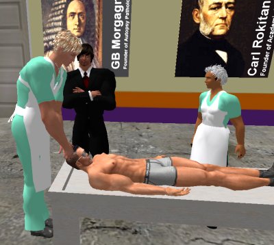
| If you have a Second Life account, please visit my teammates and me at the Medical Examiner's office. |
Teaching Pathology
 Ed's Pathology Review for USMLE I
![]()
![]()
 | Pathological Chess |
 |
Taser Video 83.4 MB 7:26 min |
 |
Click here to
see the author prove you can have fun skydiving without being world-class. Click here to see the author's friend, Dr. Ken Savage, do it right. |


 Immune Disease