Pneumonia
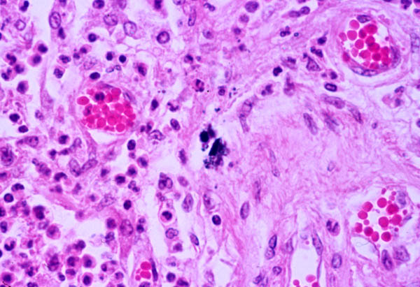
The alveoli are filled with inflammatory cells, carbon-layden type I pneumocytes are seen in the center of the field. The carbon is the black stuff in the middle of the field, possibly an old smoker. The white blood cells fighting this infection are polys (PMN's). The polys are the cells that look like clover and have dark-staining nuclei, they are located throughout the field.
The alveoli are lined by two types of epithelial cells.
TYPE I CELLS: flat cells with large cytoplasmic extensions and are the primary lining cells. These are the cells where the blood/gas exchange takes place.
TYPE II CELLS: (granular pneumocytes) are thicker and contain numerous lamellar inclusion bodies. These cells secrete surfactant.
The conversion of Type II cells to Type I cells occurs at a rate of about 1 percent per day.
Also, note the small blood vessels containing erythrocytes.
| Visitors to Tom Demark's website since Sept. 1999 |