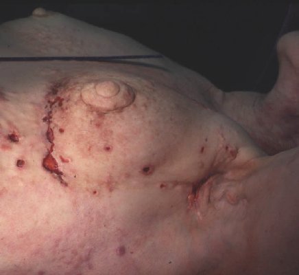
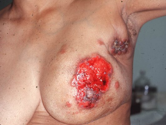
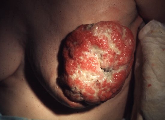
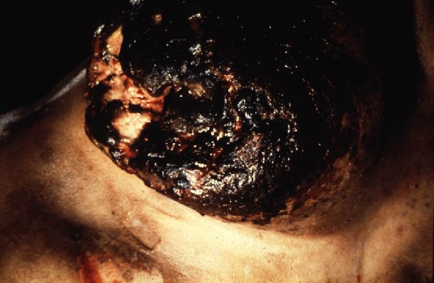
These four photos depict advanced or neglected cancers of the breast. They are not placed here to disturb or frighten you, but to emphasize the importance of early detection and treatment. You can greatly reduce the chances of this happening.
The original lump with which breast cancer presents is almost never painful. A woman who performs monthly self-examination properly, and who has mammograms as recommended, greatly reduces her risk of dying of breast cancer. Any dominant mass in the breast needs to be checked by a physician.
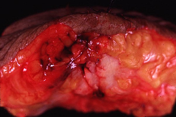
This is a typical appearance of a breast cancer. The breast has been sectioned to show the mass, which is focally hemorrhagic. The largest portion of the cancer appears as a white, infiltrating mass.

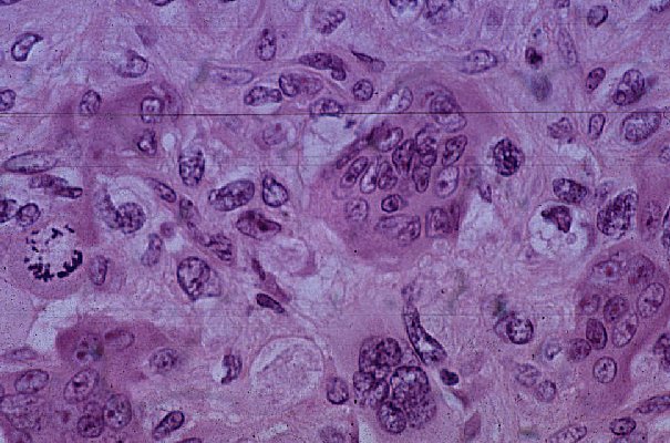
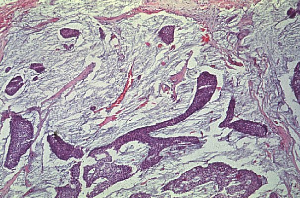
Here are some of the many histologic patterns which breast cancer can take.
The first shows lobular carcinoma, forming a few large cell groups within the lobules (purple masses). Look hard at the magnified version and you may see "Indian files" of tiny cells in the connective tissue between the glandular elements.
The second pattern is very unusual. It shows a giant cell reaction in an aggressive breast cancer.
The third pattern is a mucinous (colloid) carcinoma. The jelly-like material stains gray-blue here. These breast cancers tend to be less aggressive than most others.
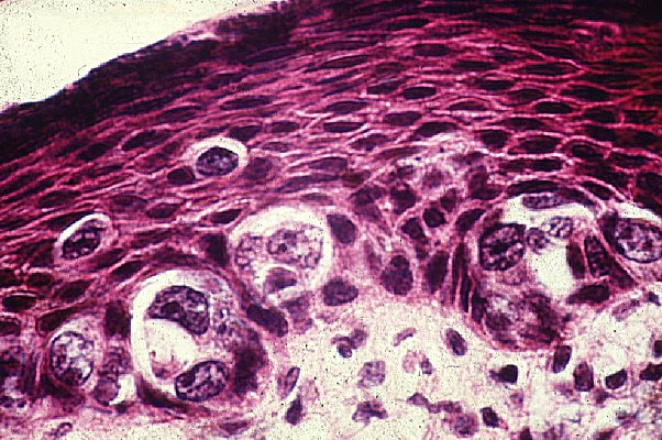
This photo shows breast cancer cells invading the stratified squamous epithelium of the nipple. This situation is called "Paget's disease of the breast." Any "rash" on the nipple is suspicious for an underlying breast cancer.
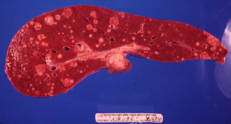
This liver contains metastatic breast cancer. Notice how the tumor nodules "umbilicate", i.e., tend to die off in their centers. Blood supply to the liver is quite variable from hour to hour, explaining this curious phenomenon. Liver metastases in breast cancer are common but seldom cause death by themselves.